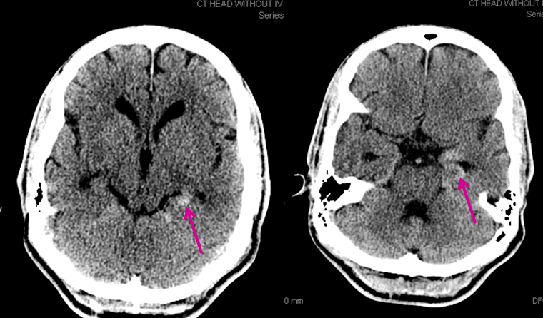
Sometimes we get a glimpse into the world of disease not usually evaluated by cerebral angiography. No doubt, this is a rediscovery of long forgotten facts from pre-cross-sectional era. This 60+ patient came in with mild confusion and amnesia. A CT scan showed hyperdensity in the posterior left hippocamal formation (pink arrows), which was initially interpreted as being in the adjacent perihippocampal cistern, raising the specter of subarachnoid hemorrhage.
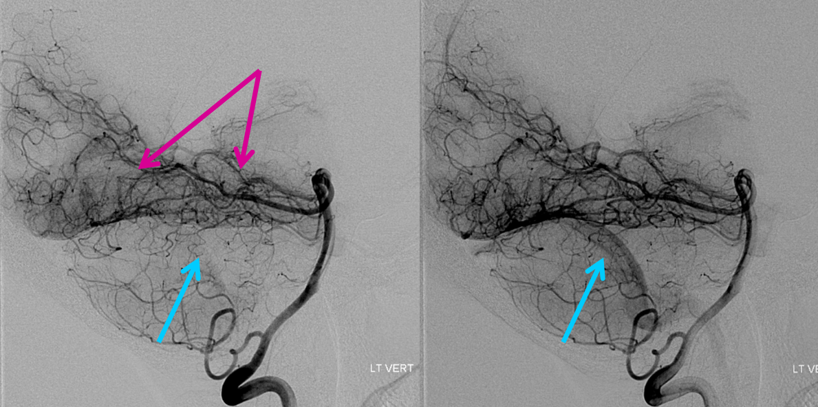
An angio was ordered. Below are sequential DSA images of the left vertebral artery injection. Look at the tremendous hyperemia in the left mesial temporo-occipital area (pink arrows), with physiologic shunting as evidenced by premature appearance of the transverse sinus (blue arrows). This is essentially the picture of parenchymal inflammation — just like any other part of the body, where inflammation makes tissues hot and hypervascular, so it is here. Of course, the differential is any kind of inflammatory/infectious process; in this clinical context, we went with encephalitis.